Fig. 4.
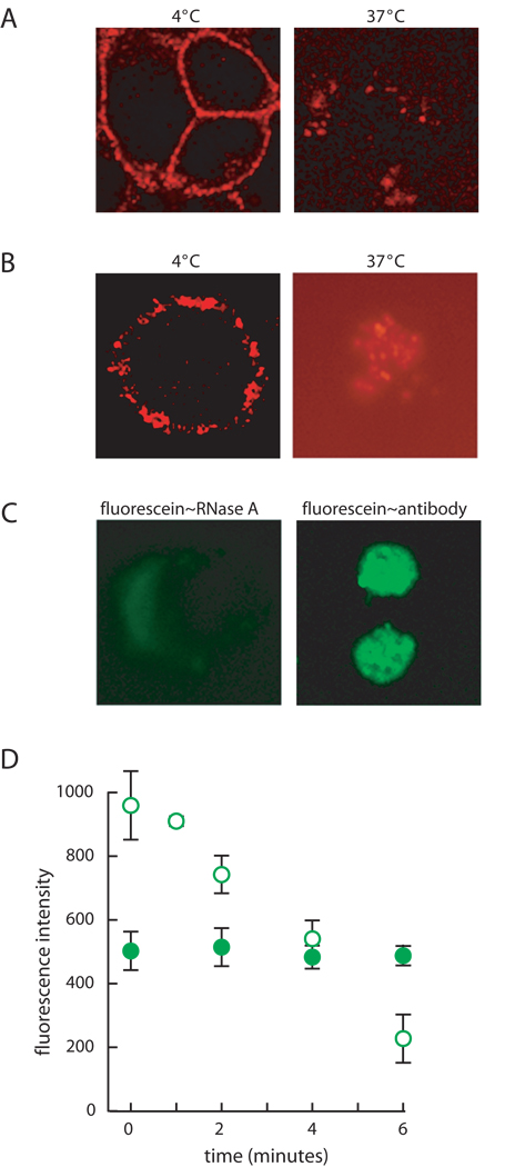
Binding and internalization of RNase A. (A) JAR cells were incubated with BODIPY–RNase A (1 µM) for 30 minutes at 4°C, and their fluorescence was visualized directly or after a 5-min incubation at 37°C. (B) K-562 cells were treated as in Panel A. (C) K-562 cells were incubated with unlabeled RNase A as described for panel A. After a 5-min incubation at 37°C, cells were visualized directly or fixed, and internalized RNase A was detected by using an appropriate primary and secondary antibody. (D) K-562 cells were incubated with fluorescein–RNase A (open; 1 µM) or OG–RNase A (filled; 1 µM) for 20 minutes at 4°C. Fluorescence intensity was measured after a 0–6-minute incubation at 37°C with a FACScan flow cytometer. Each data point represents the mean (±SE) of the fluorescence from 10,000 cells in two separate experiments.

