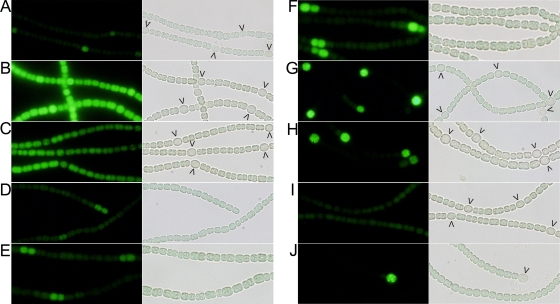
FIG. 3.
Fluorescence micrographs (left images) and light transmission micrographs (right images) of Anabaena sp. PCC 7120 bearing (A) PhetR-gfp in pSMC127, (B) the −728/−696 tsp-gfp fusion in pRR139, (C) the −184 tsp-gfp fusion in pRR151, (D to H) the −271 tsp-gfp fusion in pRR140, and (I) the −728/-696(+132) tsp-gfp fusion in pRR149 and (J) the patA mutant UHM101 bearing the −271 tsp-gfp fusion in pRR140. The images in panels A, B, C, G, I, and J were obtained 18 h after combined nitrogen was removed from the culture. The images in panels D, E, F, and H were obtained 4, 8, 12, and 24 h, respectively, after combined nitrogen was removed from the culture. The arrowheads indicate proheterocysts and heterocysts.

