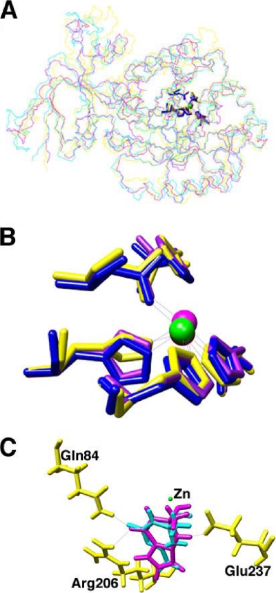
FIG. 5.
Crystal structure and docking analysis of guanine deaminases. (A) Overlay of X-ray structure backbones of guanine deaminases from B. japonicum USDA 110 (PDB 2ood; yellow), human (PDB 2uz9; blue), and C. acetobutylicum (PDB 2i9u; purple). (B) Catalytically significant residues in the guanine deaminase active sites. The spheres represent the active-site zinc atoms. (C) Docking results of guanine (purple) and ammeline (cyan) tetrahedral intermediates within B. japonicum USDA 110. Residues that interact with both substrates include Arg206, Gln84, and Glu237.

