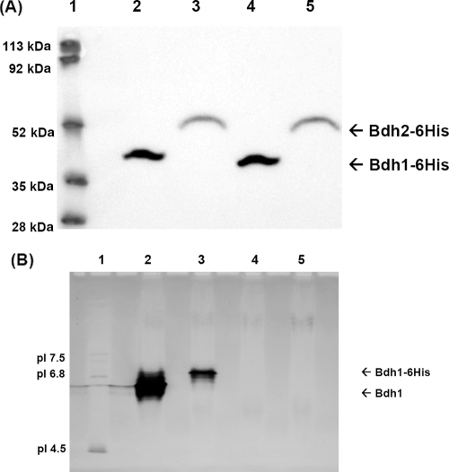
FIG. 8.
Western blot and isoelectric focusing analyses of the expression of Bdh1p-6His and Bdh2p-6His. (A) Western blot analysis. Yeast cells at an OD of ∼10 from strains WCG4-11/22a bdh1Δ(pYES2-BDH1-6His) (lane 2) and WCG4-11/22a bdh1Δ(pYES2-BDH2-6His) (lane 3) were treated as described previously (34) for Western blot analysis with an anti-His antibody. Lane 4, 54 μg of protein from extract WCG4-11/22a bdh1Δ(]pYES2-BDH1-6His); lane 5, 57 μg of protein from extract WCG4-11/22a bdh1Δ(pYES2-BDH2-6His); lane 1, molecular mass standards. (B) Isoelectric focusing analysis. Shown is an isoelectric focusing gel (pH 3 to 9) of Bdh1p, Bdh1p-6His, Bdh2p, and Bdh2p-6His visualized by butanediol dehydrogenase activity. Lane 1, pI standards; lane 2, 75 μg of protein from extract WCG4-11/22a bdh1Δ(pYES2-BDH1) containing 1.9 U of BDH activity; lane 3, 81 μg of protein from extract WCG4-11/22a bdh1Δ(pYES2-BDH1-6His) containing 0.15 U of BDH activity; lane 4, 81 μg of protein from extract WCG4-11/22a bdh1Δ(pYES2-BDH2) with no BDH activity; lane 5, 86 μg of protein from extract WCG4-11/22a bdh1Δ(pYES2-BDH2-6His) with no BDH activity.

