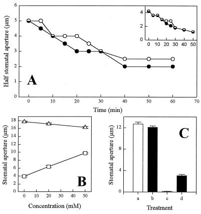
Figure 3.
Effect of inhibitors of the cADPR signaling pathway on ABA-induced stomatal closure. (A) Representative plot showing half stomatal aperture measurements after challenge of a guard cell with 1 μM ABA, where 8-NH2-cADPR had previously been loaded into the cell cytosol by pressure microinjection (○). Also shown are similar measurements for the uninjected cell from the same stoma (•). (A Inset) Half stomatal aperture measurements of a cell previously loaded with ADPR and subsequently challenged with 1 μM ABA (○) and the uninjected cell from the same stoma (•). (B) Stomatal aperture measurements from isolated epidermal strips incubated under conditions that promote stomatal opening (50 mM KCl/10 mM Mes continuously perfused with CO2-free air under constant illumination) for 2 h and subsequently incubated for a further 2 h in the same buffer containing a range of nicotinamide concentrations in the absence (▵) or presence (□) of 1 μM ABA for the final 1 h of the experiment. (C) Stomatal aperture measurements from isolated epidermal strips incubated for 3 h under conditions promoting stomatal opening in the presence or absence of inhibitor or ABA. Bar a, buffer only; bar b, 50 mM nicotinamide; bar c, 1 μM ABA; and bar d, 1 μM ABA and 50 mM nicotinamide. For B and C the data are the means ± the SEMs. A total of 120 and 160 stomatal aperture measurements for each data point were obtained for B and C, respectively.

