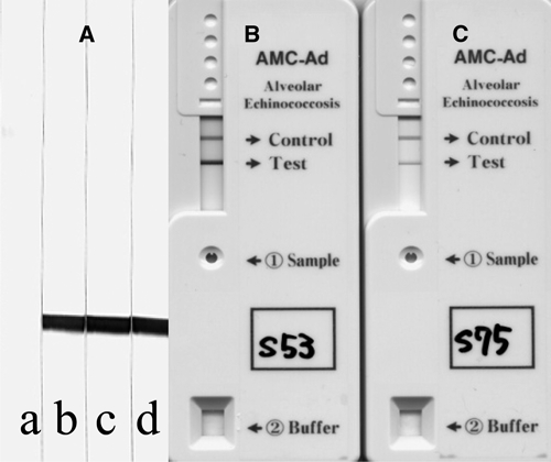
Figure 2.

Immunoblot and immunochromatographic figures of the two AE cases in Mongolia using a recombinant Em18.11,12 (A) Immunoblot figures. Lane a, shows a pooled negative control serum in 1:50 dilution, lane b, shows confirmed AE serum in 1:1000 dilution, and lanes c and d show sera from cases 4 and 5, respectively, in 1:20 dilution. (B and C) Immunochromatographic figures of cases 4 and 5, respectively.10
