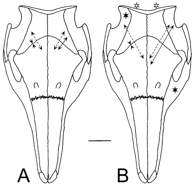
Fig. 6.

Strain in the coronal suture. Conventions as in Figure 4, data from Table 5. The scale bar represents 400 με. A: Mastication. Solid lines: late in a left side masticatory cycle when the left masseter and right temporalis are the most active muscles. Dotted lines: during the opening portion of the same cycle. Values are relative only, because a baseline could not be established during mastication. B: Muscle stimulations. The dotted lines show the result of bilateral stimulation of the neck extensor muscles (open stars). The solid lines on the left side of the figure show strain arising from the right temporalis (solid star). On the right side of the figure, the solid lines indicate strain from stimulation of the left masseter (solid star).
