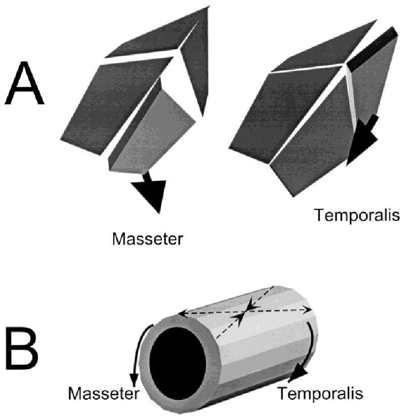
Fig. 9.

A: Suture strain as a result of muscle contraction. The masseter pulls ventrally and laterally. The resulting rotation and bending are read as tension in the coronal suture and the anterior part of the interfrontal suture. The temporalis pulls anteriorly and ventrally, causing compression in the coronal suture but tension in the interparietal. B: The muscles also torque the braincase, producing the 45° pattern reported in the frontal and parietal bones.
