
Fig. 2.
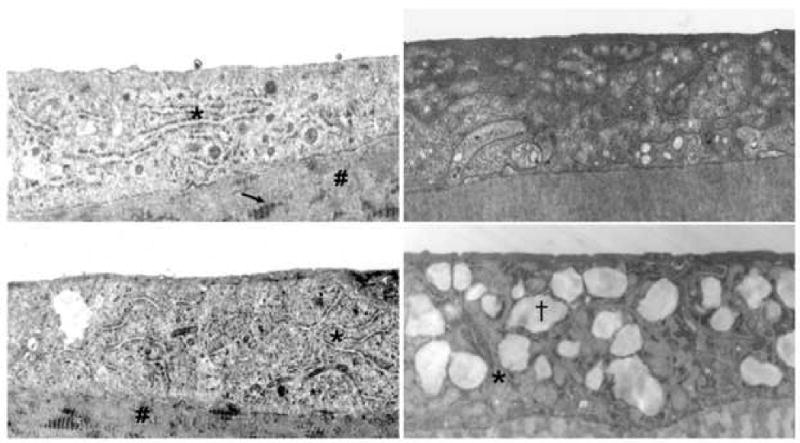
Electron microscopy images of corneal endothelium of patients with Fuchs dystrophy (upper and lower left images), granular corneal dystrophy (upper right image), and macular corneal dystrophy (lower right image). Comparison shows prominent and enlarged rough endoplasmic reticulum in the upper and lower left images as compared to the upper and lower right images. Asterisks indicate rough endoplasmic reticulum in the upper/lower left and the lower right images. # indicates posterior collagenous layer (upper/lower left) with wide-spaced collagen (arrow, upper left), typical of Fuchs dystrophy. (†) indicates intracytoplasmic vacuole typical of macular corneal dystrophy (lower right) 29. Original magnification (upper left: 15000×; lower left: 8000×; upper right: 15000×; lower right: 15000×).