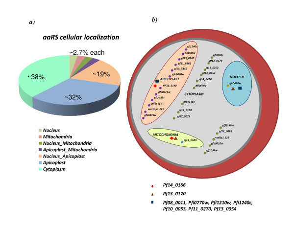
Figure 3.
(a) Percentage predicted distribution of Pf-aaRSs in different organelles within the parasite. (b) A schematic of all Pf-aaRSs and their predicted cellular localization. Detailed information regarding gene IDs can be found in additional file 1. Pf-aaRSs predicted to be common between apicoplast & mitochondria, mitochondria & nucleus and apicoplast & nucleus are marked with diamond, triangle and square shapes respectively.

