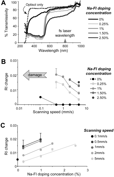
Figure 2.
Effect of Na-Fl doping on light transmissivity and RI change in living corneal tissue. (A) Light transmissivity of the preservative solution (black dotted line) and corneal wedges stored in the preservative only (black line) or in preservative doped with different concentrations of Na-Fl (gray lines). Note the strong absorption of light in the range between 300 and 500 nm in Na-Fl-doped live corneal tissues. (B) Magnitude of RI change versus scanning speed attained in corneas doped with different concentrations of Na-Fl. (C) Magnitude of RI changes attained versus Na-Fl doping concentration at different scanning speeds. Note that at each scanning speed, the RI change increased monotonically with the Na-Fl doping concentration.

