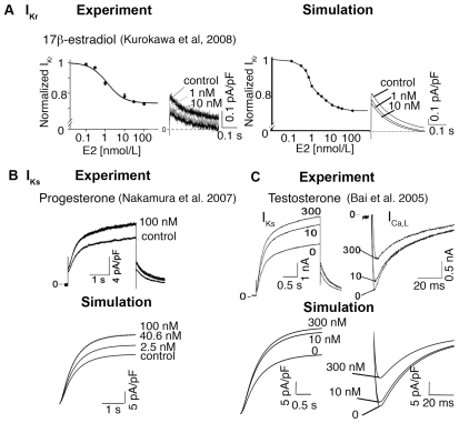
Figure 1. Effects of sex-steroid hormones E2, progesterone, and testosterone on cardiac ion channels.
(A) Dose-dependence curves are shown for experimental (left traces) and simulated (right traces) inhibition of IKr current by E2. The simulated IKr tail currents (right) compared to experimentally measured IKr (left) at −40 mV following depolarization to a test potential = +20 mV in the absence (control) or presence of E2 (1 and 10 nM). (B) IKs was experimentally recorded at a test potential of +50 mV from a holding potential of −40 mV with 0 nM and 100 nM progesterone (top traces). Simulated (lower races) IKs are shown in the presence of 0 nM (control case), 2.5 nM (follicular phase), 40.6 nM (luteal phase) and 100 nM progesterone during a voltage pulse from −40 mV to +50 mV. (C) IKs (left panels) were elicited by 3.5-s test pulses to +50 mV from a holding potential of −40 mV (experiment — top traces and simulation — lower traces) in the absence and presence of testosterone (10 nM and 300 nM). The effect on ICa,L (right panels) from experimental data (top traces) and simulated results (lower traces) during a voltage step from −40 mV to 0 mV under control condition (0 nM), 10 nM and 300 nM testosterone.

