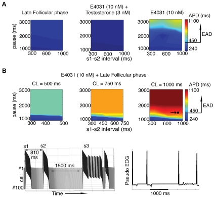
Figure 6. Pause-induced EAD susceptibility is increased in the late follicular phase of the menstrual cycle.
(A) The simulated cell was paced for 10 beats at BCL = 1000 ms (s1) followed by varying s1–s2 intervals and long pause intervals. The intervals between s1 and s2 are shown on the x-axis, pause intervals on y-axis and APD are indicated by color gradient. Simulated EAD formations under three conditions, late follicular phase (left panel), in the presence (middle) of testosterone 3 nM and E-4031 10 nM, and addition of E4031 in the absence of sex-steroid hormones (right). (B) Simulated APDs during the late follicular phase with E-4031 (10 nM) application at three basic cycle lengths (500 ms, 750 ms, and 1000 ms). The point indicated by an arrow (right panel) corresponding fiber and pseudo ECG (lower panels) under same conditions.

