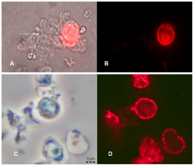
Figure 7. Effects of anidulafungin on β-1,3-D-glucan in P. carinii.
P. carinii from the lungs of immunosuppressed rats were treated with 60 µg/ml anidulafungin for 24 hrs and reacted with a mAB to β-1,3-D-glucan conjugated to Alexafluor 594. Panel A: Non-anidulafungin treated P. carinii viewed by phase microsocopy showing a cluster of trophs (arrow) attached to a cyst; Panel B: the same cluster viewed by fluorescent microscopy; no trophs were stained by the antibody; Panel C: Anidulafungin-treated P. carinii viewed by phase contrast microscopy; and Panel D: the same cluster under fluorescent excitation. Note the punctate staining pattern in both samples. All panels magnified according to the bar in Panel C.

