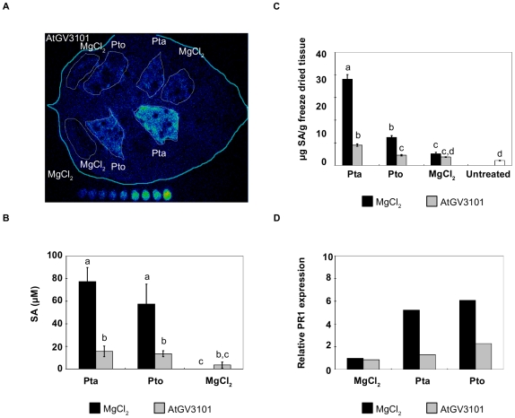
Figure 2. Agroinfiltration of N. tabacum leaves prior to P. syringae infection reduces P. syringae-elicited salicylic acid (SA) production and PR1a expression.
In all experiments, leaves were inoculated with A. tumefaciens GV3101 (AtGV3101) at 107 cfu/ml or MgCl2(AS), followed by inoculation with P. syringae pv. tabaci 11528 (Pta) or P. s. pv. tomato DC3000 (Pto) at 105 cfu/ml or with MgCl2 after 48 hours. A. The leaf shown was inoculated with AtGV3101 (top panels) and MgCl2 (bottom panels). The SA biosensor ADPWH-lux was inoculated into leaves 24 hours after infiltration with P. syringae. SA-induced luminescence was measured one hour after biosensor inoculation using a photon-counting camera. In vitro SA concentration ladders were included with each leaf imaged to allow comparison of bioluminescence between separate images. B. Numerical SA values for leaves inoculated with ADPWH-lux were calculated using a calibration curve as described in Huang et al. [26]. The chart shows average SA values from three independent experiments. GLM analysis revealed statistical differences between treatments (F = 23.6361; p<0.0001; df = 5). Means with the same letter were not significantly different at the 5% confidence level based on Tukey's HSD Test. C. SA levels in whole leaf extracts. SA content was determined by LC/MS/MS as described by Forcat et al. [61] The graph shows average values from five independent experiments. One way ANOVA revealed statistical differences between treatments (F = 24.9968; p<0.0001; df = 6). Means with the same letter were not significantly different at the 5% confidence level based on Student's t-test. Bars indicate the standard error of the mean. D. PR1a expression relative to the housekeeping gene EF1-α measured by quantitative real-time PCR. The figure shows a representative experiment. Similar ratios of PR1a expression in agroinfiltrated leaves with respect to mock-treated leaves were observed in three separate experiments.

