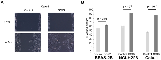
Figure 6. Consequences of Sox2 over-expression in the wound healing in vitro assay.
A. Pictures were acquired at the beginning of the experiment (t = 0, immediately after wounding) and from the same field at the end of the experiment (t = 24 h for this example from the Calu-1 cell line). B. Quantification of wound closure for the three cell lines (BEAS-2B, NCI-H226 and Calu-1). Wound sizes were measured at the beginning and end of the experiment to calculate the percentage of wound closure for control and SOX2 over-expressing cells for each of the three cell lines. SOX2 over-expression significantly stimulates cell migration compared to control cells (student's t-test).

