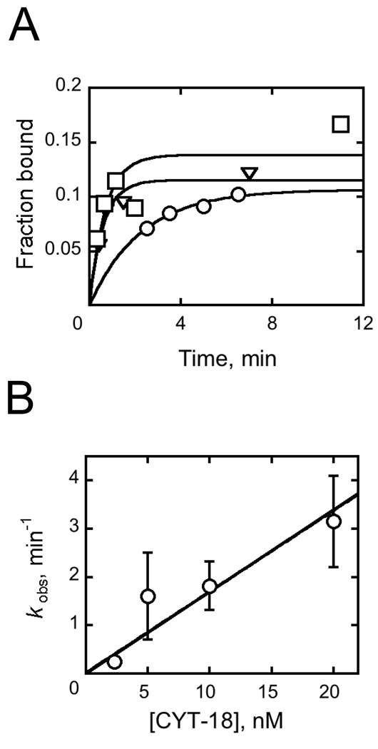
Fig. 5.
Kinetics of EΔP5abc ribozyme binding by CYT-18. A, Progress curves for radiolabeled EΔP5abc binding to CYT-18 at concentrations of 5 nM (circles), 10 nM (triangles), and 20 nM (squares) CYT-18. Aliquots were quenched with 1 µM unlabeled EΔP5abc ribozyme and then applied to filters. B, Rate constant for CYT-18 binding plotted against CYT-18 concentration. Error bars show standard deviations from two to three replicate measurements.

