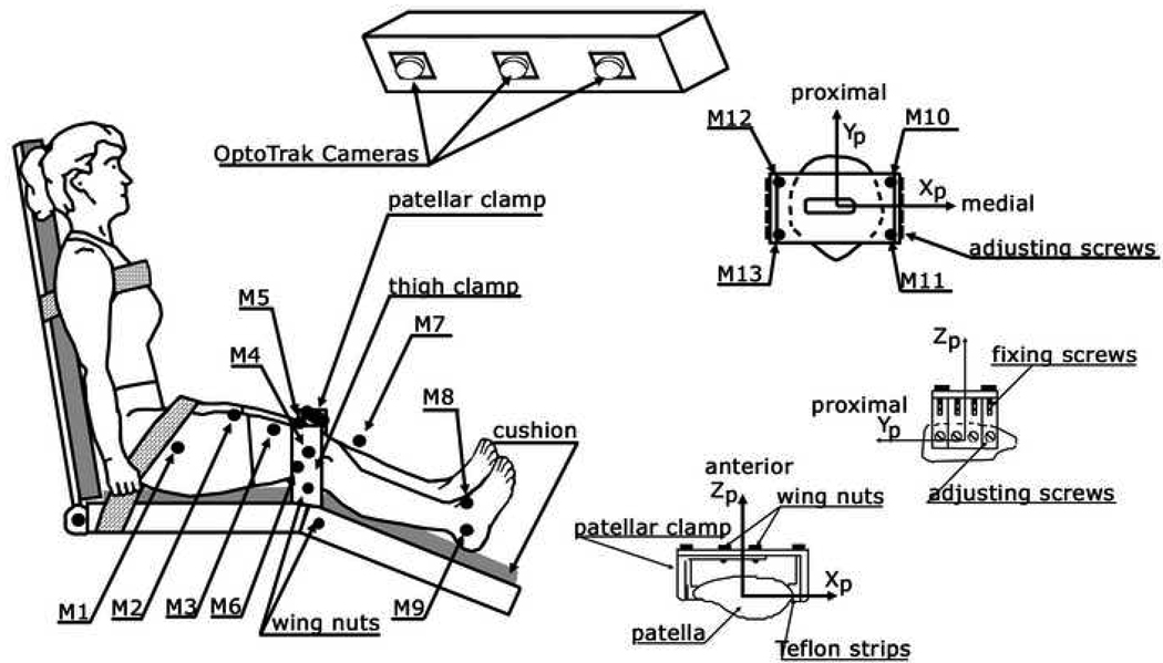
Figure 1.
Experimental setup for in vivo and noninvasive patellar tracking. The subject was seated with the thigh and trunk strapped to the seat and backrest. The femoral condyles were fixed from both the medial and lateral sides with a thigh clamp. Two pairs of wing nuts were tightened from the medial and lateral sides to fix the femoral condyles. Three markers were placed on the thigh (M1, M2, M3). Three markers were attached to the thigh clamp (M4–M6) with M4 placed on the knee flexion axis and lateral to the lateral epicondyle. Three markers were attached to the lower leg (M7–M9), and three were fixed to the four corners of the patellar clamp (M10–M13).

