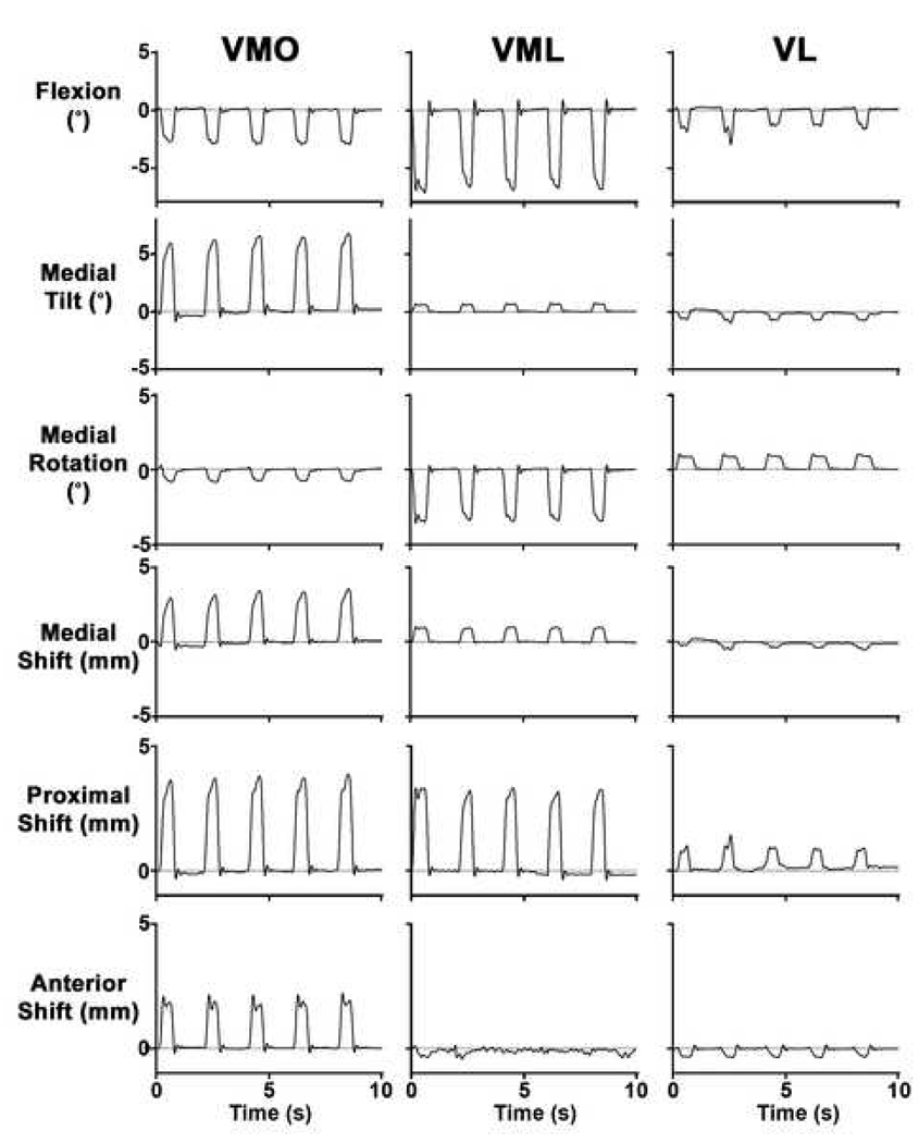
Figure 2.
Typical result for patellar tracking in healthy subjects induced through selective activation of individual quadriceps components with the knee at full extension, neutral tibial rotation, and neutral tibial abduction. The stimulation train (and thus the contraction) was repeated at 2-second intervals. From top to bottom, the six rows correspond to patellar flexion, medial tilt, medial rotation, medial shift, proximal shift, and anterior shift, respectively. The positive direction of each DOF is given for the ordinate. The zero position corresponded to the patellar position prior to stimulation.

