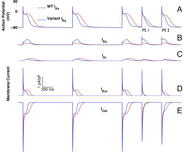
Figure 7.
Simulated double premature atrial depolarizations (400 ms coupling intervals) after a twelve-pulse pacing train (1000 ms cycle length). Data from simulations that include WT-IKs are shown in red, and those incorporating variant IKs, based on IAP54-56, are in blue (A). The first atrial premature stimulus (PS1) simulated with variant IKs was shorter than that for WT-IKs (126 vs. 193 ms). AP durations for the second premature stimulus (PS2) were 138 ms and 243 ms for variant IKs and WT-IKs, respectively. In both simulations, PS1 exhibited loss of the dome associated with membrane repolarization. However, with WT-IKs the dome was restored with PS2 while it remained absent with variant IKs. Changes in individual membrane currents are shown in (B) IKs; (C) IKr; (D) IKur; and (E) ICa-L.

