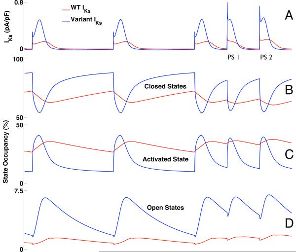
Figure 8.
IKs and channel state occupancy during the simulation of Figure 7 (1000 ms pacing train followed by double premature atrial depolarizations with 400ms coupling interval). WT-IKs is shown in red; variant IKs is in blue (A). Variant IKs exhibits much more dynamic variation in state occupancy during activation as channels transition from the closed states (B) through the activated state (C) into the open states (D). `Closed States' refers to the sum of closed states R1 and R2; `Open States' (D) refers to the sum of open states O1 and O2 (see Figure S-1A of the online data supplement).

