
Figure 1. Mechanobiology tools and their applications.
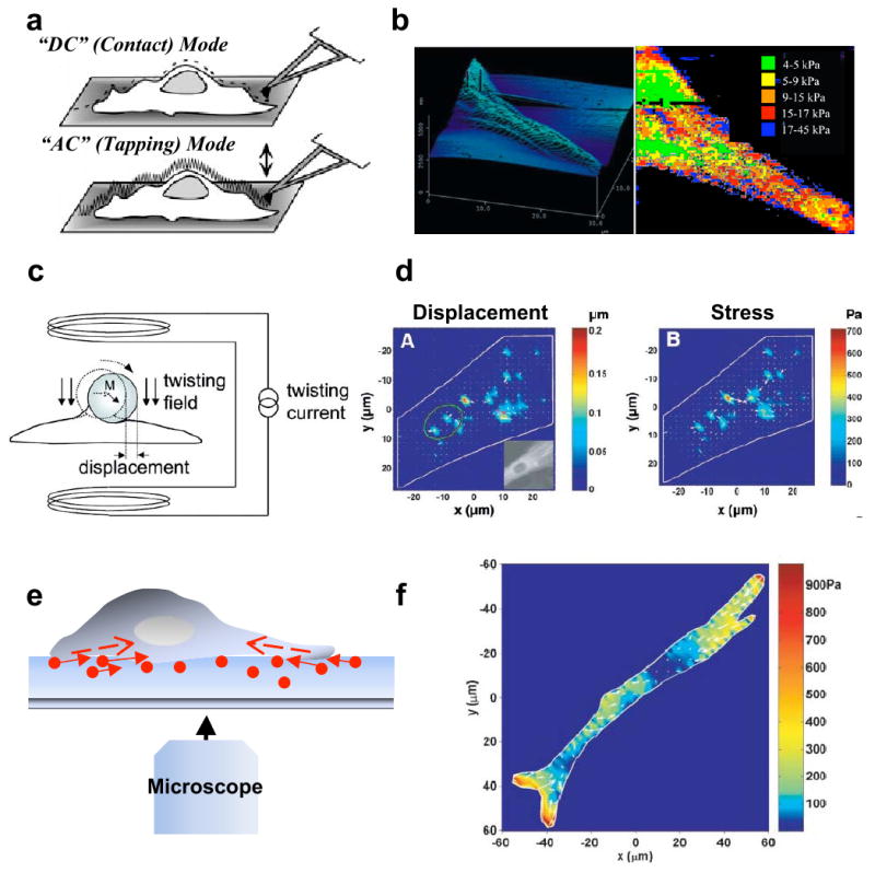
a. Atomic force microscopy (AFM). AFM can be operated either in DC (contact mode) in which the tip is brought into direct contact with the surface and the cantilever deflection is kept constant, or in AC (tapping) mode in which the tip is oscillated at its resonant frequency and a constant amplitude is maintained as the sample is scanned. Figure reprinted from [24] with permission. b. Topographic map and stiffness map of the edge of a fibroblast adhering to a fibronectin-coated glass substrate. Figure reprinted from [31] with permission. c. Magnetic twisting cytometry (MTC). In MTC, a homogeneous magnetic twisting field applied to a cell surface-adhered bead causes the bead to rotate and to displace thereby inducing shear stresses on the cell surface. Figure reprinted from [58] with permission. d. Displacement and stress maps computed using MTC for a human airway smooth muscle (HASM) cell expressing YFP-actin. Figure reprinted from [61] with permission. e. Traction force microscopy (TFM). Schematic of a cell on a hydrogel with embedded beads (circles) used in TFM. Arrows are indicative of the direction of bead motion induced by cell contractility (dotted arrows inside the cell). f. Traction field computed using TFM for a HASM cell on a 1300 Pa gel. Arrows show the direction and the relative magnitude of the tractions. Figure reprinted from [14] with permission.