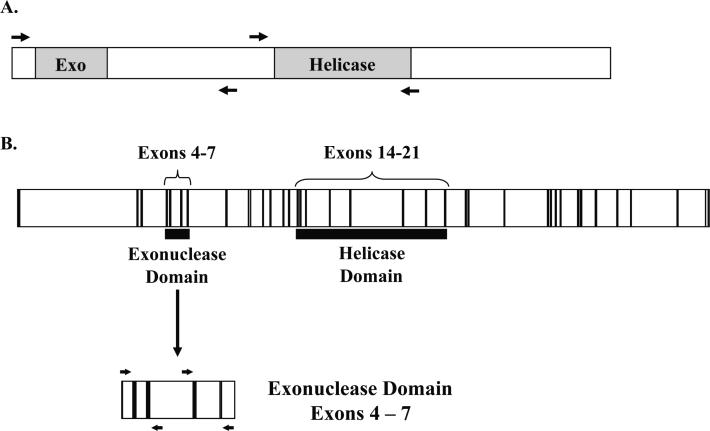
Fig. 1. Schematic of the WRN transcript and genomic WRN DNA.
(A) Regions of the WRN transcript containing the exonuclease and helicase domains (shown in grey) were amplified and sequenced, as indicated by the flanking arrows. (B) The region encoding the WRN exonuclease domain (exons 4–7) was amplified and sequenced from genomic DNA (exons are shown in black, introns shown in white). Due to large intronic regions, exons 4 and 5 were amplified together, and exons 6 and 7 were amplified together as indicated by the flanking arrows. Each exon was sequenced individually.

