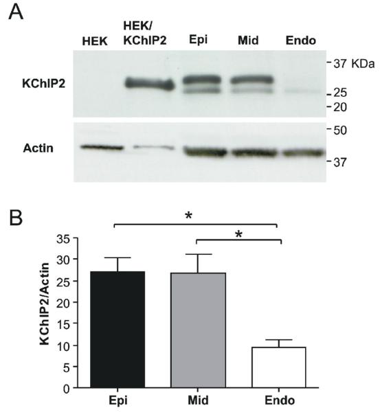
Figure 8.
A: Transmural gradient of KChIP2 protein in canine left ventricle. Representative blot of KChIP2 expression in Endo-, Mid- and Epi. KChIP2 expressing and non-transfected HEK-293 cell lysates were included as controls. For Epi, Mid and Endo, two bands sized 25-32 kDa were consistently detected for all protein isolation methods tested. The same blot was re-probed with anti-actin antibody. Panel B: Both KChIP2 bands were quantified and KChIP2 expression normalized to actin expression. Tissue from 5 dogs was analysed.

