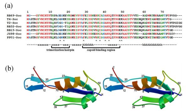
Fig. 2.
Sequence alignment and structure of Soc. (a) Alignment of Soc sequences from T4-like bacteriophages. Completely conserved residues are colored red, nearly completely conserved residues are colored green and partially conserved residues are colored blue. β-strands and α-helices are marked with an s and h, respectively. Arrow heads indicate residues that have been mutationally characterized. (b) Stereo figure showing the structure of RB69 Soc molecule as a ribbon diagram. The polypeptide is colored with rainbow colors starting with blue at the amino end.

