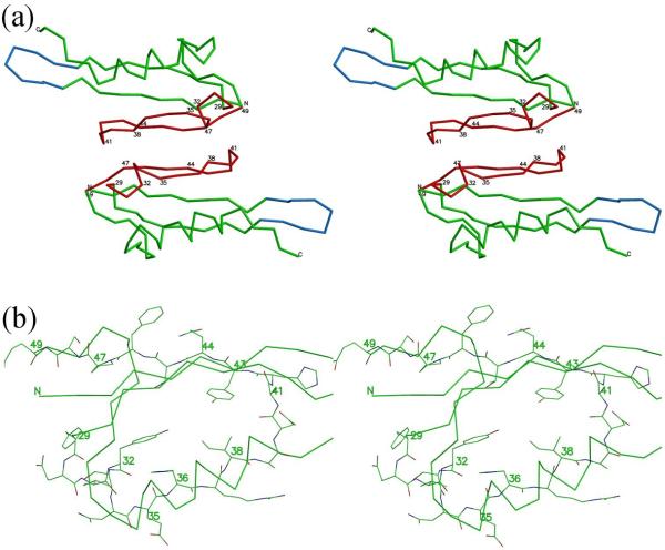
Fig. 5.
Interface of Soc molecules in the crystal corresponds to the interface between Soc and gp23 in the virus. (a) Stereo diagram showing the Cα backbone trace of two Soc molecules related by two-fold non-crystallographic symmetry. Regions of the Soc molecule that were found to interact with the gp23 capsid shell on the virus are in red. Hairpins involved in the trimerization of the Soc molecules on the capsid surface are in blue. (b) Close view of residues 29-47 involved in interactions with the capsid showing all non-hydrogen atoms. Carbon atoms are green, oxygen atoms are red and nitrogen atoms are blue. Some other parts of the structure are shown as a Cα backbone trace.

