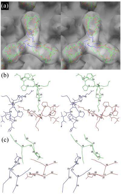
Fig. 6.
The Soc trimer. (a) Stereo diagrams of RB69 Soc fitted into the cryo-EM reconstruction of the T4 capsid, viewed down a quasi-three-fold axis. The three RB69 monomers are shown as a Cα backbone colored with the trimerization loop in blue, the gp23 binding region in red and the rest of the structure in green. (b) Stick drawing of the trimerization loop for RB69 with the three different molecules colored red, blue and green. (c) The same as B, but based on the T4 homology model showing the side chains for Lys16 and Asp18. The main chain is shown only as Cα atoms.

