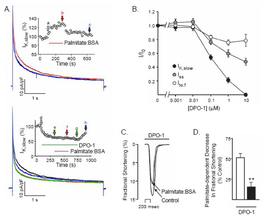
Figure 6.
Pharmacological inhibition of IK,slow by DPO-1 occludes the effects of palmitate on ventricular Kv currents and fractional shortening. (A) Representative Kv current recordings from a WT myocyte in response to depolarizing voltage steps to +40 mV from a holding potential of −70 mV, presented at 15 s intervals; currents recorded during superfusion of control solution (black), palmitate:BSA (30 µM:60 µM) (red) and following washout (blue) are displayed. IK,slow was reversibly increased by palmitate. Inset: IK,slow amplitude (normalized to control) is plotted as a function of time during the application and washout of palmitate:BSA. (B) Representative Kv currents recorded from a WT myocyte in response to depolarizing voltage steps +40 mV as described in (A) in control bath solution (black), following superfusion of 100 nM DPO-1 (green) or 100 nM DPO-1 plus palmitate:BSA (30 µM:60 µM) (red) and after washout (blue), are displayed. The addition of palmitate:BSA in the presence of DPO-1 does not measurably affect the amplitudes or the waveforms of the Kv currents. Inset: IK,slow amplitude (normalized to control) is plotted as a function of time during the application and washout of DPO-1 and DPO-1 plus palmitate:BSA. (C) Analyses of the Kv currents revealed that, in addition to inhibiting of IKslow, DPO-1 also attenuates the amplitudes of the transient (Ito,f) and steady state (Iss) Kv currents in adult mouse LVA myocytes in a dose-dependent manner. Outward Kv currents were recorded as described in the legend to Figure 4 in the presence and absence of varying concentrations of DPO-1. In each cell, the amplitudes of the individual Kv current components (Ito,f, IKslow and Iss) were determined (see Materials and Methods), normalized to the maximal current amplitude )in the same cell at each DPO-1 concentration; mean +SEM normalized values for Ito,f, IKslow and Iss are plotted as a function of DPO-1 concentration. (D) Representative single contraction record obtained from an adult WT LVA myocyte in the presence of 100 Nm DPO-1 during superfusion with control Tyrode solution containing 100 nM DPO-1 alone or 100 nM DPO-1 plus palmitate:BSA (30 µM:60 µM). In the presence of 100 nM DPO-1, the mean ± SEM decrease in fractional shortening produced by palmitate:BSA was reduced significantly (**P<0.01).

