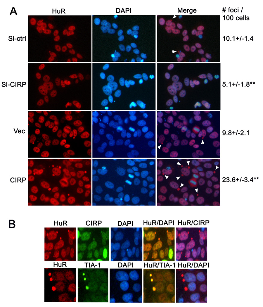
Fig. 7.
CIRP increases HuR containing cytoplasmic foci. MCF-7 cells were transfected with control siRNA (si-ctrl), CIRP siRNA (si-CIRP), pTracer-CMV2 vector (vec), or pTracer-CMV2-CIRP (CIRP). A, HuR localization was assessed using immunofluorescence analysis. Representative fluorescence micrographs of cells 72 hr after transfection are shown. HuR staining was visualized with Cy-3 conjugated secondary antibody (red). Nuclei were stained with DAPI (blue). White arrowheads in the merge panel indicate cytoplasmic foci containing HuR but not DAPI. Cytoplasmic foci containing HuR were counted in 500 cells for each condition and shown on the right as foci number per 100 cells. ** p< 0.01 as compared to Si-ctrl or Vec. B, HuR co-localization with CIRP and TIA-1 was assessed in MCF-7 cells transfected with pTracer-CMV2-CIRP using immunofluorescence analysis. Representative fluorescence micrographs of cells 72 hr after transfection are shown. HuR staining was visualized with Cy-3 conjugated secondary antibody (red). CIRP and TIA-1 were visualized with Alexa Fluor 488 (green) conjugated secondary antibody). Nuclei were stained with DAPI (blue). All experiments were repeated at least 3 times.

