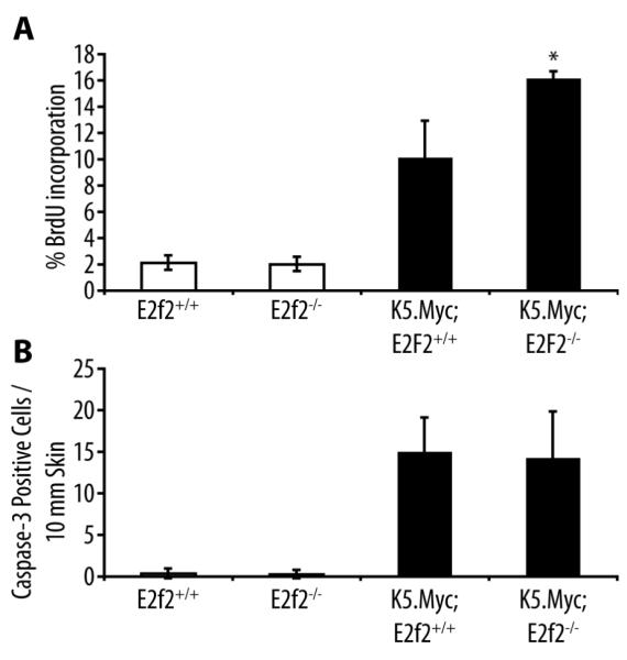
Figure 2. Inactivation of E2f2 enhances proliferation in K5.Myc transgenic epidermis.

(A) BrdU incorporation in the epidermis was analyzed by immunohistochemical staining of skin sections from wild type (E2f2+/+), E2f2−/−, K5.Myc, and K5.Myc, E2f2−/− mice. The percentage of BrdU-positive cells in the epidermis was calculated from at least four mice for each genotype. * indicates a statistically significant difference between K5.Myc, E2f2+/+ and K5.Myc, E2f2−/− mice as determined by the student’s paired t-test (p < 0.05). (B) Skin sections from the same mice used above were stained for the activated form of caspase 3 as an indicator of apoptosis. The average number of caspase 3-positive cells per 10 mm of linear epidermis was determined for each genotype.
