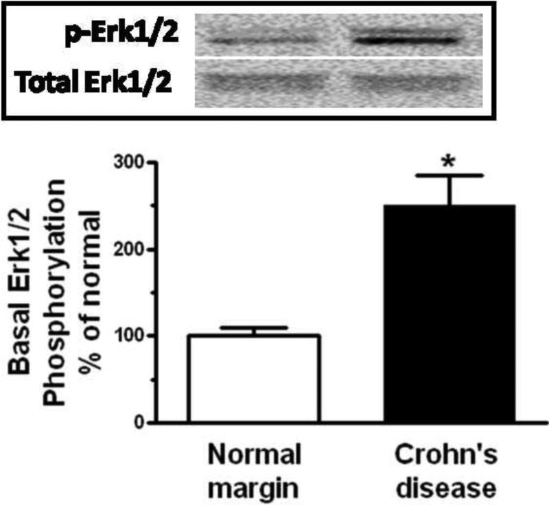
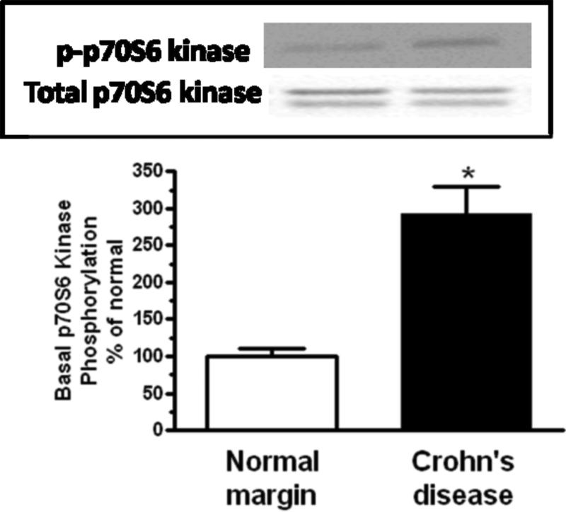
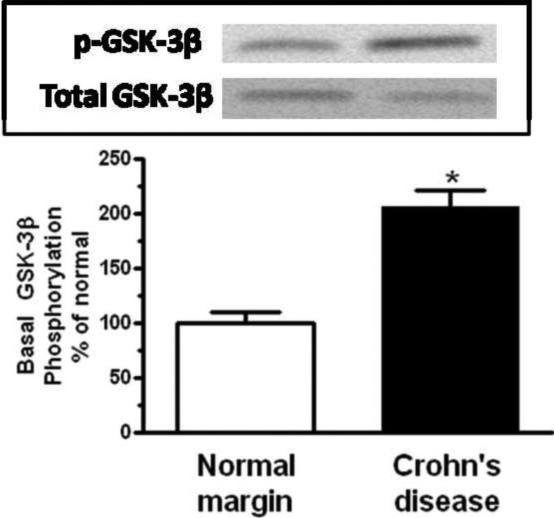
Figure 4. Basal signaling intermediate phosphorylation is increased in smooth muscle cells of stricturing Crohn's disease.
Panel A: Densitometric analysis showing increased basal Erk1/2 phosphorylation in smooth muscle cells isolated from stricturing Crohn's disease compared to normal margins. Inset: Representative immunoblots of phospho-Erk1/2(Thr202/Tyr204) and total Erk1/2 levels. Panel B: Densitometric analysis showing increased basal p70S6 kinase(Thr389) phosphorylation in smooth muscle cells isolated from stricturing Crohn's disease compared to normal margins. Inset: Representative immunoblots of phospho-p70S6 kinase(Thr389) and total p70 S6 kinase. Panel C: Densitometric analysis showing increased basal GSK-3β phosphorylation in smooth muscle cells isolated from stricturing Crohn's disease and normal margins. Inset: Representative immunoblots of phospho-GSK-3β(Ser9) and total GSK-3β. Results are expressed in relative densitometric units normalized to total signaling intermediate levels. Values represent the mean ± SE of 4 separate experiments. * denotes P < 0.05 vs normal margin.


