Fig. 7.
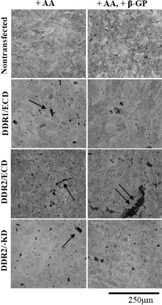
DDR ECD alters the formation of mineralized plaque in cells. Nontransfected MC3T3-E1 cells display small crystalline calcium deposits (black) throughout the sample; the size of the crystals in these samples increases slightly upon addition of the osteogenic supplement β-glycerophosphate (β-GP) to the culture media. Stable cell lines DDR1/ECD, DDR2/ECD and DDR2/-KD show increased crystal size as compared to nontransfected cells when cultured in the presence of Ascorbic acid (AA), indicated by arrows. Addition of β-glycerophosphate (β-GP) further increases the size of calcium deposits in stable cell lines DDR2/ECD and DDR2/-KD as compared to nontransfected cells. The DDR2/ECD cell line shows the most pronounced calcified crystals, indicated by double arrow. All samples were cultured for two weeks.

