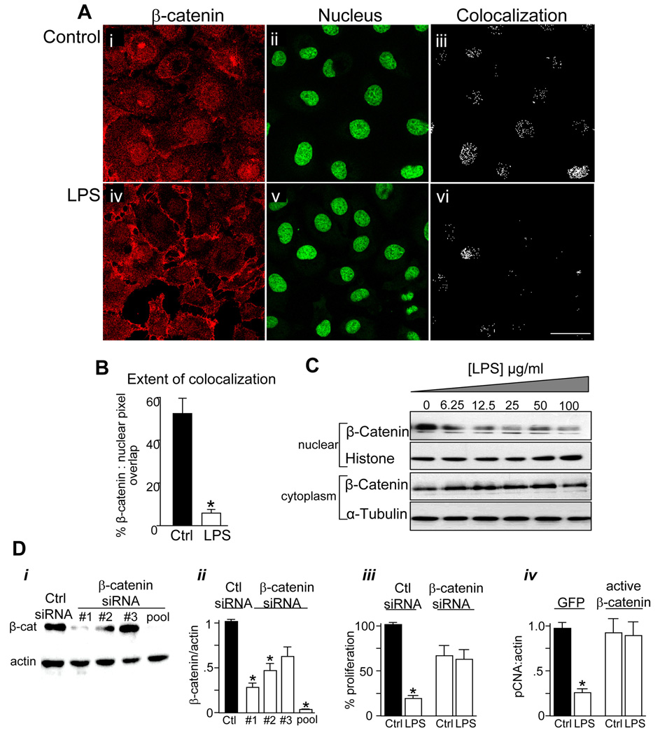
Figure 3. TLR4 activation inhibits enterocyte proliferation via inhibition of β-catenin signaling.
A: Confocal images of IEC-6 cells showing β-catenin (red, i, iv) and the nuclear marker Draq-5 (green, ii, v). Size bar=10 µm. B: Extent of colocalization (in iii and vi) was calculated using ImageJ,*p<0.05, representative of 5 separate experiments. C: SDS-PAGE of β-catenin in nuclear and cytoplasmic fractions obtained from IEC-6 cells treated with LPS as indicated, along with the nuclear marker histone and the cytoplasmic marker α-tubulin. Representative of 3 separate experiments. D: SDS-PAGE (i) and quantification of β-catenin:actin expression (ii) in IEC-6 cells transfected with siRNA to no known target (“Ctrl”) or to β-catenin. Three siRNA's (#1, #2, #3) were used either individually or in combination (“pooled”), as indicated. iii: Proliferation in IEC-6 cells treated with the indicated siRNA (XTT assay). iv: RT-PCR showing fold change in pCNA:actin in IEC-6 cells transfected with either GFP or constitutively active β-catenin and treated with LPS. *p<0.05 vs. control, representative of 3 separate experiments.

