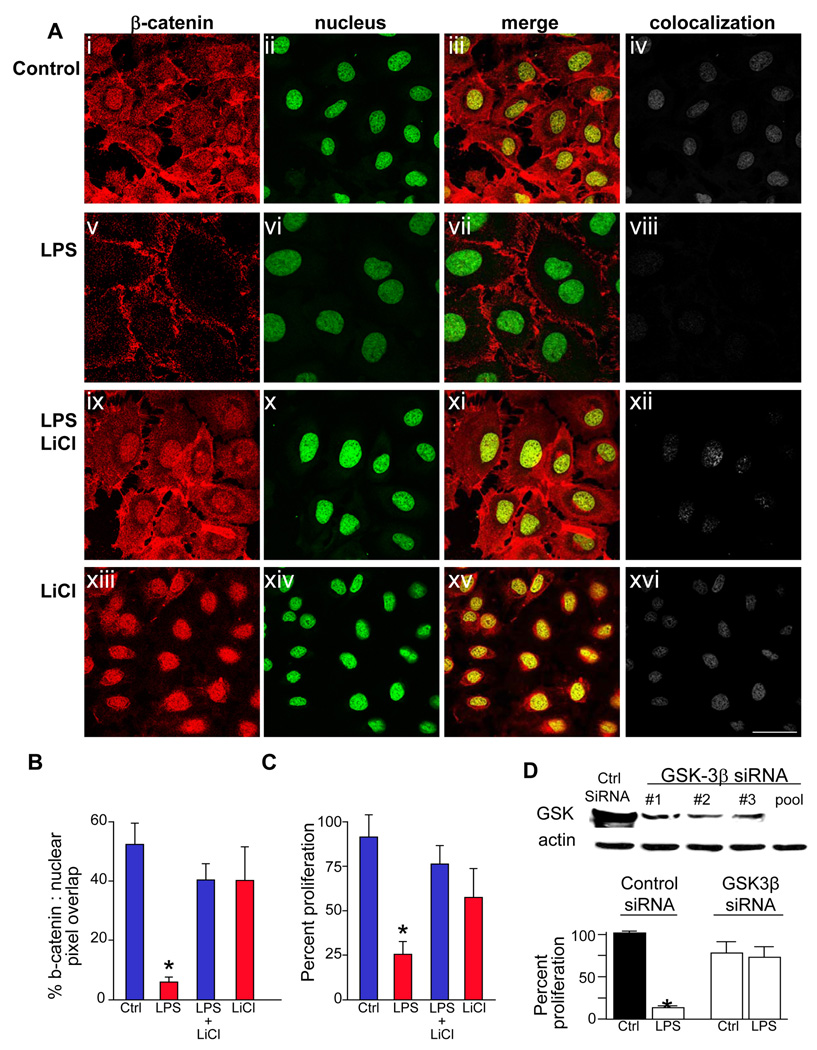
Figure 5. TLR4 activation inhibits enterocyte proliferation via GSK3β.
A: Confocal images showing immunolocalization of β-catenin (red, i, v, ix, xiii), the nuclear marker draq-5 (ii, vi, x, xiv), merged images (iii, vii, xi, xv) and colocalization of nuclear and β-catenin (iv, viii, xii, xvi) using ImageJ in IEC-6 cells treated with media (control), or the indicated treatment (LPS and/or the GSK3β inhibitor LiCl). Size bar=10µm. B: Quantification of colocalization between the nucleus and β-catenin using ImageJ.*p<0.05 vs. control cells, ANOVA. C: Proliferation of IEC-6 cells treated as indicated (XTT assay) *p<0.05 vs. control cells, ANOVA. D: upper: SDS-PAGE showing GSK3β in IEC-6 cells transfected with indicated siRNA's to GSK3β, either alone (#1,#2,#3) or in combination (“pool”). lower: proliferation of IEC-6 cells (XTT) after treatment with indicated siRNA and LPS or media (“ctrl”)*p<0.01 vs. control cells, ANOVA. Representative of 3 separate experiments.

