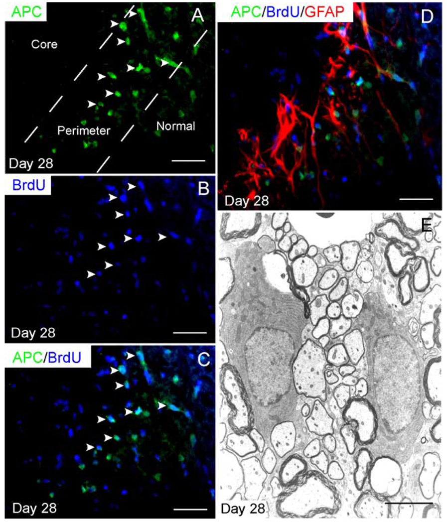
Fig. 6. Remyelinating oligodendrocytes are restricted to the perimeter of the lesion.
(A) APC+ oligodendrocytes are present in the perimeter of the lesion after 28 days, but not in the central region. Twenty-four-hour pulse labeling with BrdU at 2 dpi in animals, which were sacrificed at 28 dpi, reveals that (A–C) many APC+ oligodendrocytes in the perimeter of the lesion are BrdU+. (D) Triple label immunohistochemistry reveals that new oligodendrocytes are closely associated with the band of reactive astrocytes in the perimeter of the lesion. (E) Ultrastructural analysis reveals that oligodendrocytes in the perimeter of the lesion produce thin myelin, characteristic for CNS-type remyelination; these may be distinguished from the thicker, spared myelin. Scale bar = 25 µm in (A– C); 28 µm in (D); and 6 µm in (E).

