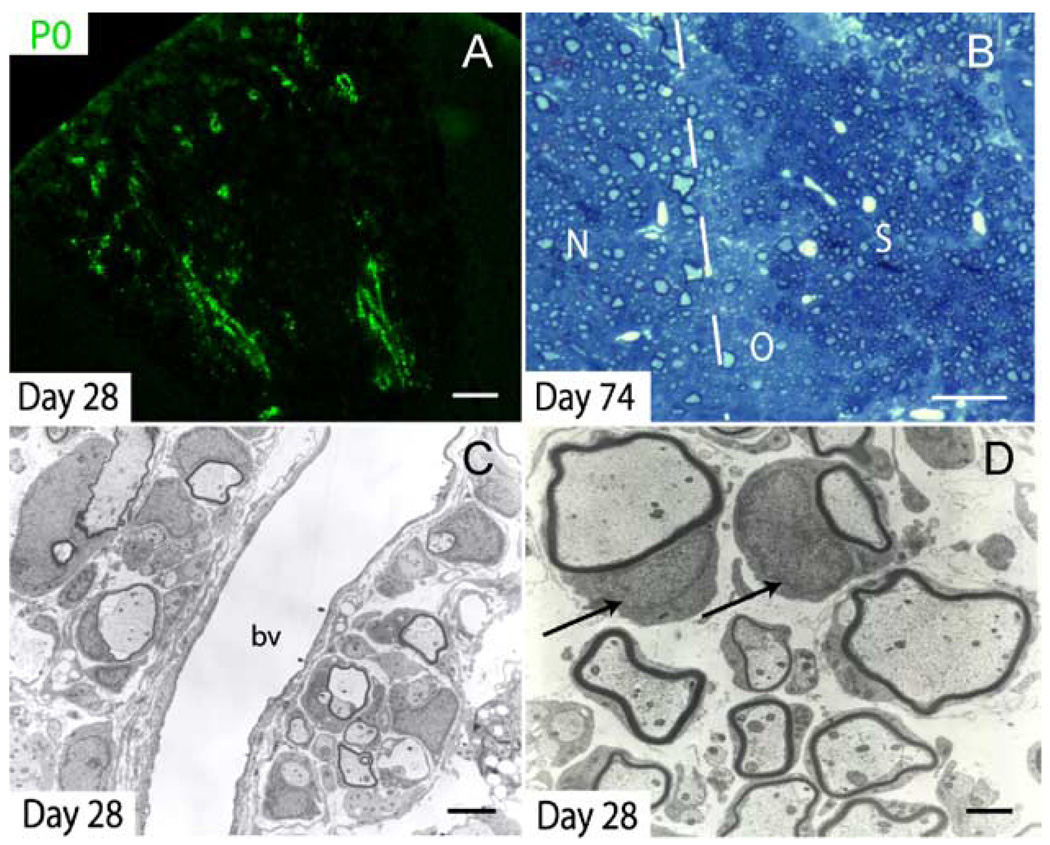
Fig. 7. Schwann cell remyelination in the lesion core.
(A) P0 immunohistochemistry demonstrates considerable Schwann cell-mediated remyelination as early as 28 dpi. Schwann cell remyelination begins in a distinct perivascular pattern. (B) After 74 days, Schwann cell (S) remyelination is extensive in the central portion of the lesion while oligodendrocyte (O) remyelination is limited to the perimeter of the lesion, adjacent to normal (N), uninjured white matter. In toluidine blue-stained plastic sections, Schwann cell myelin appears darker than CNS myelin. (C) Ultrastructural analysis confirmed the presence of characteristic Schwann cell remyelination in the central core of the VLF at 28 dpi; bv = blood vessel. (D) Higher magnification of remyelinating Schwann cells in the center of Day 28 lesions. Schwann cell nuclei are indicated by arrows. Scale bar = 90 µm in (A); 30 µm in (B); 3 µm in (C); and 1 µm in (D).

