
Fig. 3.
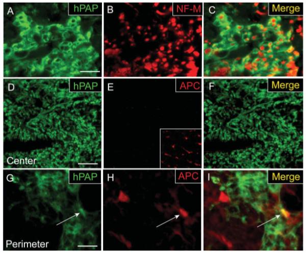
Adult OPCs engrafted into EB-X lesions ensheath axons but do not differentiate into mature oligodendrocytes in the center of lesions. hPAP immunohistochemistry for engrafted cells 4 weeks after transplantation demonstrates that transplanted cells exhibit a characteristic ring-like morphology and many of these rings ensheath NF-M+ axons. (A–C) Despite their myelin-like formations around axons, double-label immunohistochemistry for hPAP and the mature oligodendrocyte marker APC/CC-1 shows that engrafted cells in the center of EB-X lesions do not differentiate into oligodendrocytes (D–F). Inset in (E) shows positive APC/CC-1 staining for oligodendrocytes in the uninjured, contralateral VLF. An occasional hPAP+/APC+ cell (white arrow) was observed in the perimeter of such lesions (G–I). Scale bar = 20 μm (A–C);36 μm in (D–F); 16 μm in (G–I).