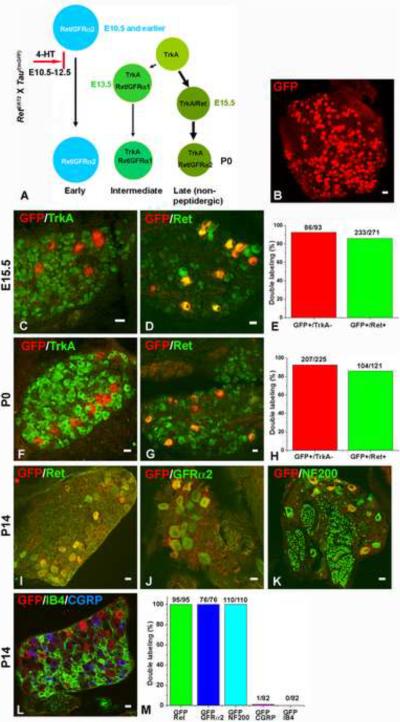
Figure 2. Genetic Labeling of the early Ret+ DRG neurons.
A. Outline of the chemical-genetic strategy for labeling the early Ret+ neurons. RetERT2 and Tauf(mGFP) mice were crossed, and timed pregnant mothers were gavaged with 1.0 to 1.5 mg 4-HT per day from E10.5 to E12.5. B. Whole-mount anti-GFP immunostaining of a labeled L5 DRG. 280 ± 25 GFP+ neurons were labeled. Eight L5 DRGs from four animals of two litters were examined. C–D: Double immunostaining of GFP with TrkA (C) or Ret (D) in E15.5 labeled DRGs. 86/93 (GFP+/TrkA− neurons / total GFP+ neurons) GFP+ neurons are TrkA−, and 233/271 (GFP+/Ret+ neurons / total GFP+ neurons) GFP+ neurons are Ret+. Lumbar DRGs, n=6 from three litters. 4–6 sections from each animal were quantified and shown in E. F–G: Double immunostaining of GFP with TrkA (F) or Ret (G) in P0 labeled DRGs. 207/225 (92%) GFP+ neurons are TrkA−, and 104/121 (86%) GFP+ neurons are Ret+. Lumbar DRGs, n=7 from four litters for GFP/TrkA staining, and n=4 from two litters for GFP/Ret staining. Quantifications are shown in H. I–L: Double immunostaining of GFP and Ret (I), GFP and GFRα2 (J), GFP and NF200 (K), and GFP and CGRP and IB4 in P14 labeled DRGs. All GFP+ neurons (95/95) are Ret+ at this time (Note that, due to the fixation conditions needed for this experiment, Ret immunoreactivity is detected only in those DRG neurons expressing a high level of Ret protein). In addition, GFP+ neurons are GFRα2+ (76/76) and NF200+ (110/110), but not CGRP+ (1/82) or IB4+ (0/82). Quantifications are shown in M. Lumbar DRGs, n=4 from two litters. Scale bar for panel B: 50μm, other panels: 20μm

