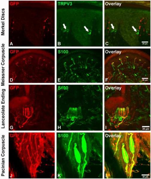
Figure 3. The early Ret+ DRG neurons are the RA mechanoreceptors associated with Meissner corpuscles, Pacinian corpuscles and longitudinal lanceolate endings.
A–C: GFP+ fibers do not innervate Merkel cells in the footpad (339/356 {95.2%} of Merkel cells, labeled with the TrpV3 antibody, are not associated with GFP+ axons). The locations of Merkel cells are indicated by white arrows. D–F: GFP+ fibers innervate Meissner corpuscles, visualized by S100 immunostaining, in dermal papillae (114/172 {66.3%} Meissner corpuscles are innervated by GFP+ fibers). G–I: GFP+ fibers form longitudinal lanceolate endings associated with hair follicles, shown by S100 immunostaining (78/184 {42.3%} of hair follicles are associated with GFP+ longitudinal lanceolate endings). J–L: Whole-mount anti-GFP and anti-S100 staining of the periosteum membrane of the fibula. Note that a single GFP+ fiber innervates each Pacinian corpuscle (55/60 {91.7%} of Pacinian corpuscles are innervated by GFP+ fibers). Immunostainings were performed using P14 RetERT2;Tauf(mGFP) mice treated with 4-HT from E10.5 and E12.5. Quantifications were made using four P14 RetERT2;Tauf(mGFP) mice from two litters.

