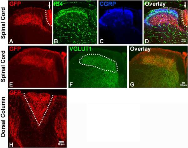
Figure 4. Central projections of early Ret+ DRG neurons innervate layers III through V of the spinal cord.
A–D: Central projections of GFP labeled early Ret+ DRG neurons in the upper lumbar level of the spinal cord. Note the presence of GFP+ fibers in both lamina III through V of the spinal cord and the dorsal column (arrow indicates the area which is also outlined by the dotted line). Nociceptor axons innervating spinal cord layers I and II were visualized with CGRP immunostaining (blue) and IB4 binding (green), respectively. E–G: Double staining of GFP and VGLUT1. VGLUT1 labels synapses of the central projections of mechanoreceptors (white dotted line) and proprioceptors (Hughes et al., 2004; Oliveira et al., 2003). H: Dorsal column of the cervical spinal cord. Note that while GFP+ fibers are present in both the gracile (V shape tract, inside the white dotted line) and cuneate fasiculi (area outside the white dotted line), they are greatly enriched in the gracile fasiculus. N=4 from two litters for P14 RetERT2;Tauf(mGFP) mice treated with 4-HT from E10.5 to E12.5.

