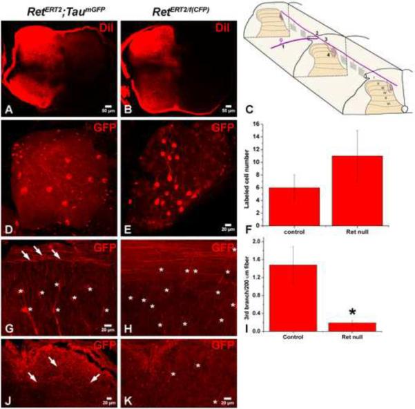
Figure 8. Ret is required autonomously for the formation of the 3rd branch of central RA mechanosensory axons.
A–B: DiI labeling of thoracic DRGs of E15.5 RetERT2;Tauf(mGFP) and RetERT/f(CFP) mice. Consistent with published findings (Ozaki and Snider, 1997), mechanoreceptors have already extended central axonal projections to spinal cord layers III through V at E15.5. C: Illustration of RA mechanoreceptor central projections, which can be subdivided into four steps. This illustration is adapted from (Brown, 1981). D: Whole mount anti-GFP staining of a control RetERT2;Tauf(mGFP) DRG from an animal treated with 0.6mg of 4-HT. Note that the 1st order central projections are visible in the DRG. On average, 6 ± 2 GFP+ neurons are labeled per thoracic DRG (12 DRGs in total, N=3 from two separate litters). E: Whole mount anti-GFP staining of a RetERT/f(CFP) DRG from an animal treated with 1mg of 4-HT. Note that 1st order central projections are visible in the DRG. On average, 11 ± 4 CFP+ neurons are labeled per thoracic DRG (20 DRGs in total, N=4 from three separate litters). F: Quantification of labeled DRG neuron number. G: Anti-GFP staining with sagittal thoracic spinal cord sections of RetERT2;Tauf(mGFP) mice. Note that the 3rd order axonal branches (white arrows) originate from rostral-caudal running fibers and penetrate the spinal cord. “*” indicates the position of blood vessels which are autofluorescent and seen in mice of both genotypes. H: Anti-GFP staining of sagittal thoracic spinal cord sections of RetERT/f(CFP) mice. Note that very few 3rd order axonal branches originating from rostral-caudal running fibers are found in these mice. I: Quantification of the numer of 3rd order axonal branches for the two genotypes. On average, 1.48 ± 0.41 3rd order axonal branches are observed in every 200μm rostral-caudal fiber in RetERT2;Tauf(mGFP) control mice (N=3 from two litters, and more than 100 rostralcaudal fibers were measured) while 0.19 ± 0.05 3rd order axonal branches are observed in every 200μm rostral-caudal fiber in RetERT/f(CFP) mutant mice (N=4 from three litters, and more than 100 rostral-caudal fibers were measured). J–K: Anti-GFP staining using horizontal lumbar spinal cord sections taken from RetERT2;Tauf(mGFP) (J) and RetERT/f(CFP) (K) mice. Similar to that observed using sagittal thoracic sections, there are several GFP+ central projections in RetERT2;Tauf(mGFP) control mice at this age, but almost no CFP+ central projections are found in the RetERT/f(CFP) mice.

