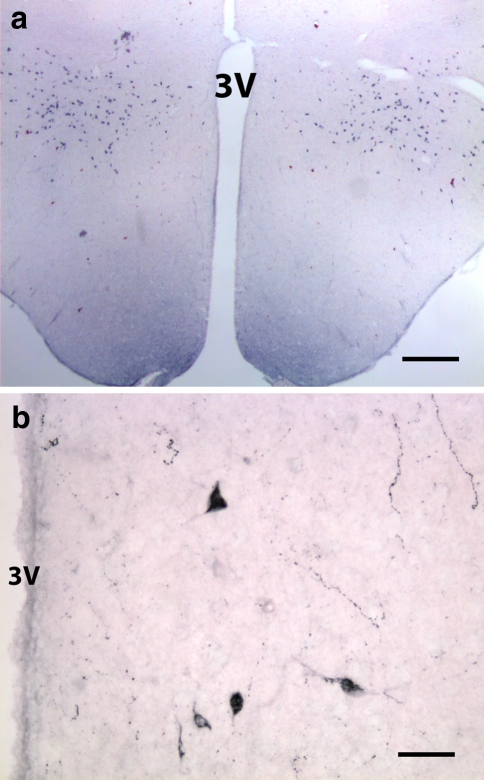
Fig. 1.
Coronal section through the hypothalamus of adult rat labeled with orexin-B antiserum. a A low magnification showing orexin-B-positive neurons distributed bilaterally in the lateral hypothalamic area. b Higher magnification of a section immunolabeled for Orexin-A. A few positive neurons are also seen in the dorsal and dorsomedial hypothalamic area, near the third ventricle (3 V). Scale bars: a 400 μm; b 50 μm

