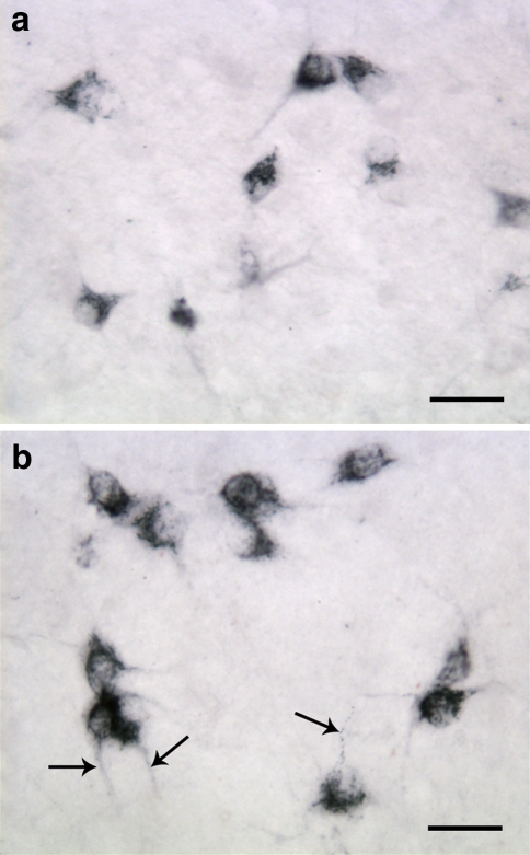
Fig. 3.
Frontal sections of the hypothalamus of 2-week-old rat processed for immunohistochemical detection of orexin-A (a) and orexin-B (b). The size of the orexinergic neurons is bigger compared to those in 1-week-old animals (see Fig. 2), and the neurites are better developed (arrows). The granular immunoreaction product is more evenly distributed in the cytoplasm. Scale bars: 30 μm

