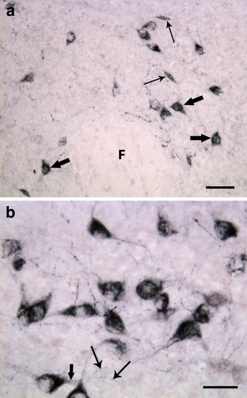
Fig. 4.
Close-up of the lateral hypothalamus of adult rat. a Orexin-A-immunoreactive neuronal pericarya and varicose neuronal fibers located in the perifornical area. The cell somata are well developed and two major types of neurons are well distinguishable: spindle-shaped neurons (thin arrows) with two main neurites arising from the opposite poles of the cell body, and multipolar neurons (thick arrows) with several major neurites emerging from a stellate-shaped soma. F, fornix. (b) Higher magnification of orexin-B positive neurons. A primary dendrite is indicated with thick arrow, and thin arrows are pointing to two secondary arborizations. Scale bars: a 50 μm; b 30 μm

