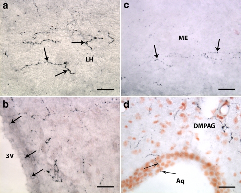
Fig. 5.
Orexin-A and -B expressing neuronal fibers projecting to different parts of the brain of adult rat. a A dense network of orexin-A containing axonal varicosities (arrows) in the LH. b Micrograph illustrating the proximity of orexin-A positive fibers to the ventricular system. Some of the neuronal processes (arrows) protrude between the ependymal cells lining the third ventricle (3 V). c Moderate density of orexin-B immunolabeled projections (arrows) to the external part of the median eminence (ME). d Orexin-A reactive fibers at the level of the mesencephalon. Moderate density of the labeled varicosities, some of which protrude into the lumen of the cerebral aqueduct (Aq) (arrows). DMPAG, dorsomedial periaqueductal gray. Scale bars: 30 μm

