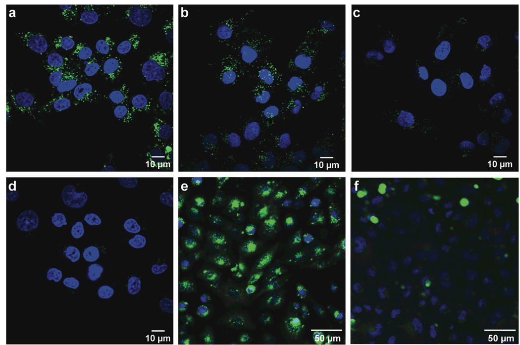
Figure 2.
Images of the internalization of GFP variants into living human and rodent cells. HeLa cells were incubated with cpGFP (a, 10 µM; b, 1 µM; c, 0.1 µM) and eGFP (d, 10 µM) for 3 h in Opti-MEM medium at 37 °C. Cells were then placed in fresh medium for 1 h and stained with Hoescht 33342 (blue) and propidium iodide (red) for 15 min prior to visualization by confocal microscopy. (e) CHO-K1 and (f) CHO-745 cells (which are GAG-deficient) were incubated with cpGFP (2 µM) for 3 h at 37 °C in Opti-MEM medium. Cells were then placed in fresh medium for 1 h and stained with Hoescht 33342 (blue) and propidium iodide (red) for 15 min prior to visualization.

