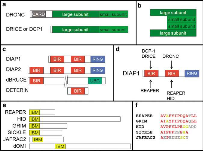
Figure 2.
Domain structure of apoptotic proteins in Drosophila. Not drawn to scale. (a) Schematic outline of the zymogen form of the initiator caspase, DRONC, and the effector caspases, DRICE and DCP-1. Initiator caspases such as DRONC contain a long prodomain that harbors regulatory motifs such as the caspase activation and recruitment domain (CARD). Effector caspases have only short prodomains. (b) After cleavage of the zymogen form of effector caspases, two large and two small subunits form the active caspase. (c) Schematic outline of IAPs in Drosophila. BIR, Baculovirus IAP repeat; RING, really interesting new gene; UBC, ubiquitin conjugation. (d) Binding preferences of the two BIR domains of DIAP1. (e) The RHG proteins in Drosophila. IBM, IAP-binding motif. (f) Alignment of the IBM of several RHG proteins. Residues in red are identical and in yellow are conserved

