
Fig. 6.
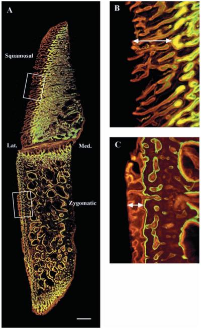
Frontal section of a zygomatic arch viewed under ultraviolet light (see Fig. 2 for orientation). (A) Low-power view showing the squamosal and zygomatic bones separated by the horizontal part of the zygomatico-squamosal suture. Note that more bone is labeled (lighter regions) laterally (Lat.) than medially (Med.), and that the tissue is organized differently in the two bones. The squamosal bone consists of numerous thin, mediolaterally oriented laminae, whereas the zygomatic bone has large randomly oriented spaces in the central region, surrounded by a more compact outer rim. (B,C) Enlargements of the lateral borders of the squamosal (B) and zygomatic (C) bones, as indicated by the boxes in A. In the zygomatic bone (C), the double labels (red and green) mostly parallel the bone surface, whereas in the squamosal bone (B), the labels are oriented around the mediolateral laminae. The arrows indicate the distance between tne calcein-labeled region and the edge of the bone surface. This was used as an index of bone growth. Scale bar, 1 mm.