
Fig. 7.
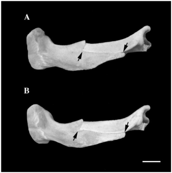
Photograph of lateral views of pig zygomatic arches from the zygomatic flange growth study. (A) Two months after removal of the masseter fibers that attach to the flange (see Fig. 1A). (B) Control sibling (masseter intact). The distance between the arrows denotes the length of the zygomatic flange as measured by the length of the horizontal portion of the zygomatico-squamosal suture. Note that both flanges are of comparable length. Scale bar, 1 cm.