
Fig. 8.
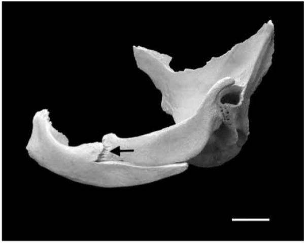
Photograph of a pig zygomatic arch in lateral view. The squamosal and zygomatic bones have been partly disarticulated to show the vertical component of the zygomatico-squamosal suture (arrow). Note that the squamosal portion of this suture lies medial to the zygomatic portion. Scale bar, 1 cm.